Which epithelial type is highlighted? This captivating question sets the stage for an enthralling narrative, offering readers a glimpse into a story that is rich in detail and brimming with originality from the outset. As we delve into the intricacies of histological staining techniques, we will uncover the secrets behind identifying and visualizing the diverse array of epithelial tissues that form the foundation of our bodies.
Epithelial tissues, with their remarkable diversity in structure and function, play a pivotal role in maintaining homeostasis, protecting against external threats, and facilitating a multitude of physiological processes. Understanding the techniques used to highlight specific epithelial cell types is essential for unraveling the mysteries of human biology and advancing our knowledge of disease mechanisms.
Epithelial Tissue Classification
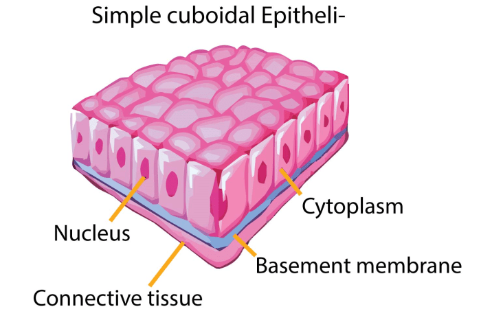
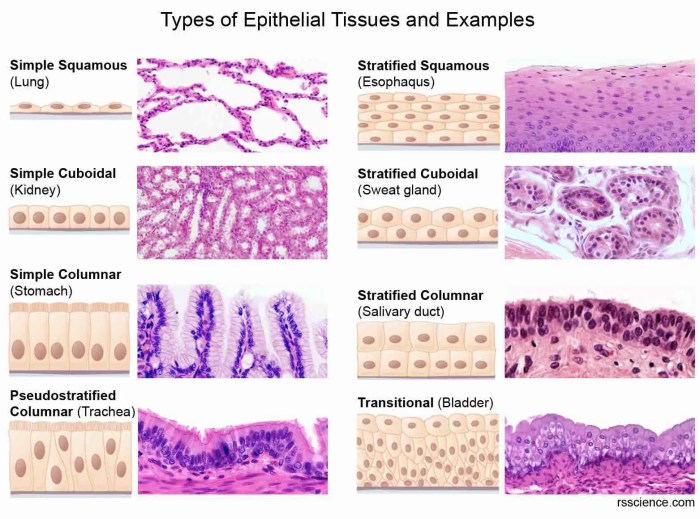
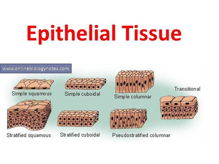
Epithelial tissue, the lining of organs and body cavities, is classified based on the shape and arrangement of its cells. This classification system helps describe the structure and function of different types of epithelium.
Cell Shape
- Squamous:Thin, flattened cells, resembling scales.
- Cuboidal:Cube-shaped cells with a central nucleus.
- Columnar:Tall, column-shaped cells with a nucleus near the base.
Arrangement
- Simple:A single layer of cells.
- Stratified:Multiple layers of cells, with the basal layer attached to the basement membrane.
- Pseudostratified:Cells appear to be stratified, but only the basal cells reach the basement membrane.
Types of Epithelium
| Type | Cell Shape | Arrangement | Location |
|---|---|---|---|
| Simple Squamous | Squamous | Simple | Capillaries, alveoli |
| Simple Cuboidal | Cuboidal | Simple | Kidney tubules, salivary glands |
| Simple Columnar | Columnar | Simple | Intestine, stomach |
| Stratified Squamous | Squamous | Stratified | Skin, esophagus |
| Stratified Cuboidal | Cuboidal | Stratified | Sweat glands |
| Stratified Columnar | Columnar | Stratified | Rare, e.g., pharynx |
| Pseudostratified Columnar | Columnar | Pseudostratified | Trachea, nasal cavity |
Histological Staining Techniques: Which Epithelial Type Is Highlighted

Histological staining techniques are essential for visualizing and studying epithelial tissues. These techniques involve the use of dyes and stains to highlight specific cellular components and structures, allowing researchers to identify and characterize different types of epithelial cells and their organization within the tissue.
Hematoxylin and Eosin (H&E) Staining
H&E staining is one of the most commonly used histological staining techniques. It utilizes two dyes: hematoxylin, which stains the nuclei of cells blue, and eosin, which stains the cytoplasm pink. This staining method provides a basic overview of tissue structure and allows for the identification of different cell types based on their nuclear and cytoplasmic characteristics.
- Advantages:Simple and cost-effective; provides good contrast between nuclei and cytoplasm.
- Disadvantages:Can lack specificity for certain cell types; may not reveal fine cellular details.
Immunohistochemistry (IHC)
IHC is a technique that uses antibodies to specifically label and visualize target proteins within cells or tissues. Antibodies are highly specific and can be used to detect the presence and localization of specific proteins, including those that are characteristic of different epithelial cell types.
- Advantages:Highly specific and sensitive; allows for the identification and localization of specific proteins.
- Disadvantages:Can be more expensive and time-consuming than other staining techniques; requires careful optimization of antibody concentrations.
Periodic Acid-Schiff (PAS) Staining
PAS staining is a technique that highlights the presence of carbohydrates, particularly glycoproteins and glycogen, within cells and tissues. It is often used to visualize the basement membrane, which is a specialized extracellular matrix that separates epithelial cells from the underlying connective tissue.
- Advantages:Specifically stains carbohydrates; useful for identifying basement membranes and mucus-producing cells.
- Disadvantages:Can lack specificity for certain types of carbohydrates; may not provide detailed cellular information.
Transmission Electron Microscopy (TEM)
TEM is a technique that uses a beam of electrons to generate high-resolution images of cells and tissues. It allows for the visualization of ultrastructural details, including the organization of organelles and the cell membrane. TEM can be used to study the fine structure of epithelial cells and their interactions with neighboring cells and the extracellular matrix.
- Advantages:Provides extremely high-resolution images; allows for the visualization of ultrastructural details.
- Disadvantages:Requires specialized equipment and expertise; can be time-consuming and expensive.
Immunohistochemistry

Immunohistochemistry (IHC) is a powerful technique used to identify and localize specific proteins within cells and tissues. It utilizes antibodies, which are highly specific molecules that bind to target proteins of interest.
In the context of epithelial tissues, IHC can be employed to characterize and differentiate various cell types based on their unique protein expression profiles. Antibodies targeting specific epithelial markers, such as cytokeratins, mucins, and adhesion molecules, can be used to identify and distinguish different types of epithelial cells.
Applications of Immunohistochemistry
Immunohistochemistry finds wide applications in both research and diagnostic settings. In research, IHC helps elucidate the molecular mechanisms underlying epithelial development, differentiation, and disease progression. It enables researchers to study the expression and localization of specific proteins in different epithelial cell types and investigate their role in various physiological and pathological processes.
In diagnostics, IHC is a valuable tool for identifying and characterizing epithelial neoplasms. It aids in tumor classification, grading, and prognosis, guiding appropriate treatment decisions. IHC can also detect minimal residual disease, assess response to therapy, and identify potential therapeutic targets.
Electron Microscopy
Electron microscopy is a powerful imaging technique that uses a beam of electrons to create detailed images of biological specimens. It offers much higher resolution than light microscopy, allowing scientists to visualize ultrastructural details of cells and tissues.
Types of Electron Microscopy
- Transmission Electron Microscopy (TEM):A beam of electrons passes through the specimen, allowing visualization of internal structures and organelles.
- Scanning Electron Microscopy (SEM):A beam of electrons scans the surface of the specimen, providing three-dimensional images of the topography.
- Scanning Transmission Electron Microscopy (STEM):Combines features of TEM and SEM, providing both internal and surface information at high resolution.
Applications in Studying Epithelial Tissues
Electron microscopy has played a crucial role in advancing our understanding of epithelial biology:
- Cell Structure:Visualizes the detailed architecture of epithelial cells, including cell membranes, organelles, and intercellular junctions.
- Tissue Organization:Reveals the arrangement of epithelial cells within tissues, including cell polarity, basement membrane, and cell-cell interactions.
- Cellular Dynamics:Captures dynamic processes such as cell division, migration, and secretion, providing insights into tissue homeostasis and repair.
Examples of Advancements, Which epithelial type is highlighted
Electron microscopy has led to significant breakthroughs in epithelial biology:
- Identification of Tight Junctions:Revealed the presence of tight junctions between epithelial cells, which play a critical role in maintaining tissue integrity.
- Characterization of Microvilli:Visualized the finger-like projections on the apical surface of intestinal epithelial cells, which increase surface area for nutrient absorption.
- Discovery of Exocytosis and Endocytosis:Captured images of vesicles involved in cellular transport processes, providing evidence for these fundamental mechanisms.
Functional Significance of Epithelial Cell Types

Epithelial cells exhibit remarkable diversity in their structure and organization, reflecting their specialized functions in different organs and tissues. Their functional significance is directly linked to their unique cellular architecture and the interactions they establish with the underlying connective tissue and the external environment.
Protection
Epithelial cells play a crucial role in protecting underlying tissues from mechanical damage, dehydration, and invasion by pathogens. Keratinized stratified squamous epithelium, found in the epidermis of the skin, provides a tough and impermeable barrier against abrasion and desiccation. The mucous membranes lining the respiratory, digestive, and urogenital tracts secrete mucus, which traps and removes foreign particles and microorganisms.
Homeostasis
Epithelial cells contribute to maintaining homeostasis by regulating the exchange of substances between the body and the external environment. The simple squamous epithelium of the alveoli in the lungs facilitates the diffusion of oxygen and carbon dioxide, while the cuboidal epithelium of the proximal convoluted tubules in the kidneys regulates the reabsorption and secretion of ions and solutes.
Absorption
Specialized epithelial cells in the small intestine, known as enterocytes, possess microvilli that greatly increase the surface area for the absorption of nutrients. These cells actively transport nutrients from the lumen into the bloodstream, facilitating the body’s nutritional requirements.
Secretion
Epithelial cells in glands secrete various substances, including hormones, enzymes, and mucus. The exocrine pancreas secretes digestive enzymes into the duodenum, while the endocrine pancreas secretes hormones like insulin and glucagon into the bloodstream.
Sensation
Certain epithelial cells, such as taste buds and olfactory cells, are specialized for sensory reception. Taste buds contain taste cells that detect different chemical stimuli, while olfactory cells in the nasal cavity detect odor molecules.
Epithelial-Mesenchymal Transition
Epithelial-mesenchymal transition (EMT) is a reversible process in which epithelial cells lose their epithelial characteristics and acquire a mesenchymal phenotype. This process plays a crucial role in embryonic development, tissue remodeling, and disease progression, including cancer.EMT involves a series of molecular and cellular changes.
Epithelial cells lose their cell-cell adhesion molecules, such as E-cadherin, and gain expression of mesenchymal markers, such as vimentin and N-cadherin. They also undergo cytoskeletal reorganization, becoming more motile and invasive.
EMT in Cancer Progression and Metastasis
EMT has been implicated in cancer progression and metastasis. In early stages of cancer, EMT can promote tumor cell invasion and migration, allowing them to escape the primary tumor and enter the bloodstream or lymphatic system. Once in circulation, these cells can seed distant organs, leading to the formation of metastases.EMT
can also contribute to resistance to cancer therapy. Mesenchymal cells are more resistant to radiation and chemotherapy than epithelial cells, making it more difficult to treat advanced cancers.
Q&A
What are the key advantages of using histological staining techniques?
Histological staining techniques offer several key advantages, including their ability to provide high-resolution images of tissue samples, allowing for detailed examination of cellular morphology and organization. Additionally, these techniques enable researchers to selectively highlight specific tissue components, such as epithelial cells, by utilizing dyes or antibodies that bind to specific molecular targets.
How can immunohistochemistry be used to identify specific epithelial cell types?
Immunohistochemistry is a powerful technique that utilizes antibodies to target and visualize specific proteins within cells. By using antibodies that bind to epithelial-specific markers, researchers can identify and characterize different epithelial cell types within a tissue sample. This technique is particularly useful for studying the distribution and expression of specific proteins in epithelial tissues.
What is the role of electron microscopy in studying epithelial tissues?
Electron microscopy provides an ultrastructural view of epithelial tissues, allowing researchers to examine the fine details of cellular architecture and organization. This technique offers unparalleled resolution, enabling the visualization of cellular components such as organelles, membranes, and cytoskeletal elements. Electron microscopy has been instrumental in advancing our understanding of epithelial cell function and differentiation.